Researchers will no longer have to choose between studying a single human brain as a patchwork of fragmented images or a distant, pixelated view of large structures.
A new imaging platform developed by a US team instead seamlessly combines the finer details of brain cells, their connections and contents, with maps of entire networks of neurons throughout the brain that hold up the overall architecture. of the brain.
These elements of brain biology exist at enormously different scales, from the nanometer-sized gaps of synapses to centimeter-long regions of the brain, which until now have required multiple samples from multiple brains to analyze using different technologies on different platforms.
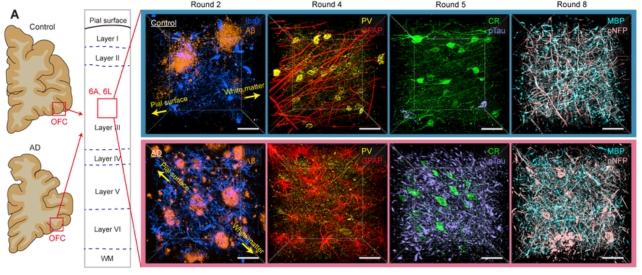
In the first demonstrated use in human tissue, imaging two whole brains, the platform has detected distinct changes in the brain of a person with Alzheimer’s disease.
The new platform includes three essential elements to slice, process and then image brain tissue with “unprecedented resolution and speed,” according to the research team that developed it, led by Kwanghun Chung, a chemical engineer at the Institute of Technology in Massachusetts (MIT). .
First, an innovative device cuts brain tissue into sections. It uses carefully tuned vibrations to avoid abrasion, cutting cells cleanly like ultra-thin slices without dislocating their connections.
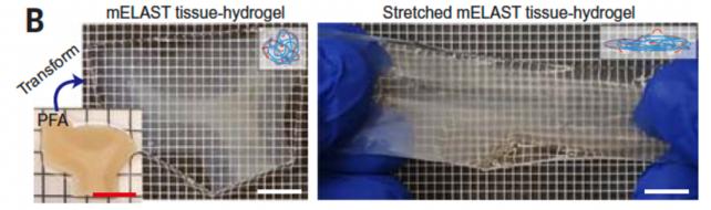
A chemical technique then reversibly transforms those tissue sections into a stretchable, expandable tissue-hydrogel, ripe for antibody labeling and high-resolution imaging of proteins and other internals.
Finally, a computational tool ‘stitches’ the cut tissues back together and maps the connections between individual cells. Those ‘projections’ of single brain cells can then be integrated with profiles that capture the molecules expressed in each cell.
“We need to be able to see all these different functional components—the cells, their morphology and their connectivity, their subcellular architectures and their individual synaptic connections—ideally within the same brain” to be able to compare the whole brain and find individual differences, he says. Chung.
“This technology pipeline really enables us to extract all these important features from the same brain in a fully integrated way.”
The tissue-turned hydrogel gently inflates tissue sections so they can be clearly imaged; and a pump stably infuses tissue with fluorescent dyes to produce consistent stains across whole organs.
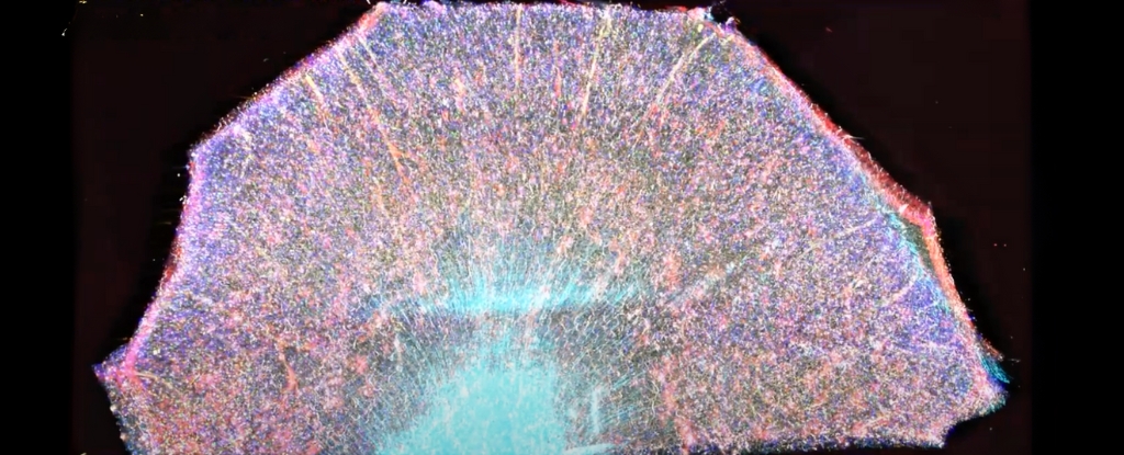
In a stunning display of the platform’s imaging capabilities, the researchers provide examples where they labeled an entire hemisphere of the brain, then zoomed in to get a picture of cell circuits, followed by single cells and their connections through junctions called synapses.
frameborder=”0″ allow=”accelerometer; Play automatically; clipboard-write; encrypted media; gyroscope; picture-in-picture; web-share” referrerpolicy=”strict-origin-when-cross-origin” allowfullscreen>
As for how the platform reconstructs those connections across multiple tissue sections, the computer tool has an algorithm that matches blood vessels exiting one layer and entering an adjacent one, and traces the extensions of neighboring neurons, called axons.
Putting it all together, the researchers imaged the entire brains of two generous donors, one with Alzheimer’s disease and the other without.
They detected the common pathological features of Alzheimer’s, including the accumulation of amyloid plaques and tau tangles, and the shrinkage of brain cells, but their imaging also captured some finer differences.
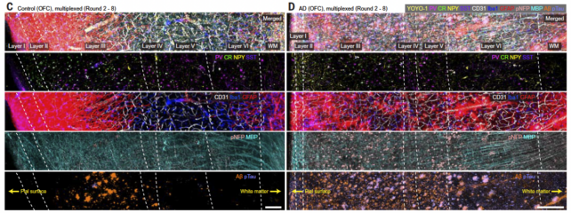
The axons of the brain cells in the Alzheimer’s patient were swollen. Brain cells in regions loaded with tau and amyloid proteins had also lost their protective myelin sheath and withdrawn from their neighbors.
This “supports neuroimaging studies that suggest severe impairments in orbitofrontal cortex connectivity in the late stages of Alzheimer’s disease,” the team wrote in their paper.
However, this gallery represents only a snapshot in time of only two brains.
frameborder=”0″ allow=”accelerometer; Play automatically; clipboard-write; encrypted media; gyroscope; picture-in-picture; web-share” referrerpolicy=”strict-origin-when-cross-origin” allowfullscreen>
Scientists have produced some incredibly detailed images of the human brain recently, zooming in on one cubic millimeter of brain tissue — a decade-long effort that ultimately produced 1.4 petabytes of data.
Imaging how the brain changes as it slowly degenerates into diseases like Alzheimer’s is a somewhat more difficult task because researchers often work with postmortem brain tissue donated at the end of someone’s life, or by relying on traditional whole-brain scans like MRI, hoping. to detect changes before the onset of the disease.
It’s also not yet clear how the platform can adapt to the rapidly unfolding advances in brain imaging, but the team is optimistic that their system will help drive the development of new therapies and maximize the amount of information extracted from valuable donor tissues.
“This pipeline allows us to have almost unlimited access to tissue,” says Chung. “We can always go back and look at something new.”
The study was published in Science.